Vein Specialists of the Carolina’s studies veins and associated diseases. We treat both the Deep Venous system and the superficial system.
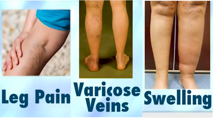
Most vein practices only treat the superficial system of veins. The only thing that needs treatment in the superficial system is reflux. Reflux is leaking one way valves that let the blood flow backwards into the legs. This can occur when standing or sitting. This is okay, bu it completely ignores the other causes of vein disease. Other venous problems include obstruction of the veins and ineffective pumping action of the calf muscle. Another vein condition completely ignored is the outflow of the leg veins into the pelvis.
Obstruction can occur from a blood clot in the deep veins (DVT). If the clot is in the legs, deep Vein Thrombosis (DVT) can be detected on the leg ultrasound. The clot however could be in the pelvic veins that the legs drain into. If the clot is in the pelvic veins it will not be detected by the leg ultrasound because it doesn’t image higher than the groin. The other cause of venous obstruction in the pelvis is compression of the veins by other structures. This was once thought to be rare because our methods of detecting it were very insensitive. Results would often show to be normal despite the obstruction really being there.
At Vein Specialists, we study the deep system in the legs and the pelvis with two additional tests that most practices do not. One is called an APG, Air Plethysmography. The other is a pelvic venous ultrasound.
Veins Disease – Air Plethysmography:
APG is a test that measures venous volume in the leg with a very low pressure cuff around the calf. The patient is studied lying down, with the leg elevated, standing and while performing tip toe maneuvers. Before ultrasound came along this was the only way to study veins unless a venogram or dye study was performed. The dye study involved an IV, leg tourniquets and lots of x-ray time. This dye can cause kidney damage or allergic reactions. The APG can be done in the office with none of those risks. The APG did not just detect the presence or absence of reflux or clot. Additionally it can quantitate the severity of it. It also studied the calf muscle pump’s ability to eject blood from the leg.
Veins Disease – Pelvic Venous Ultrasound:
Ultrasound proves to be equally if not more effective at fining blood clots. It does not involve IV’s, Dye or radiation. It is now the gold standard. Later it became effective at identifying reflux. But this study, which is the one nearly all vein specialists use today, only detects the presence or absence of reflux, not its severity. It does not study the outflow restrictions that may exist or the ability of the calf to pump blood out of the leg. Unfortunately, the APG also got forgotten with arrival of the ultrasound. One reason is APG is a fairly meticulous test and great care and technique have to be used to get consistent results. One of the world’s experts in venous disease, Dr Seshadri Raju of Jackson Mississippi, said “the study of vein disease has devolved with the lack of use of the APG”.
The pelvic venous ultrasound, although sounding simple is very difficult to learn. It is not even taught in ultrasound school. The world’s experts in developing the techniques have only been doing it about five years. We have been developing our skills for the last two years.
Our commercials say “all we do is veins, all day, every day” we also mean: we do ALL veins, all day, every day.